Arch Iran Med. 28(9):536-537.
doi: 10.34172/aim.34777
Photoclinic
A Diagnostic Triad in the Vesicular Stage of Incontinentia Pigmenti
Thien Nguyen Conceptualization, Data curation, Formal analysis, Investigation, Methodology, Software, Visualization, Writing – original draft, 1, * 
Tuan Anh Vu Funding acquisition, Project administration, Resources, Supervision, Validation, Writing – review & editing, 2
Author information:
1Emergency Resuscitation Department, Quy Hoa National Dermatology Hospital, South Quy Nhon, Gia Lai, Vietnam
2Board of Directors, Quy Hoa National Dermatology Hospital, South Quy Nhon, Gia Lai, Vietnam
Copyright and License Information
© 2025 The Author(s).
This is an open-access article distributed under the terms of the Creative Commons Attribution License (
https://creativecommons.org/licenses/by/4.0), which permits unrestricted use, distribution, and reproduction in any medium, provided the original work is properly cited.
Cite this article as: Nguyen T, Vu TA. A Diagnostic Triad in the Vesicular Stage of Incontinentia Pigmenti. Arch Iran Med. 2025;28(9):536-537. doi: 10.34172/aim.34777
A 2-month-old female infant presented with cutaneous symptoms since birth, characterized by vesicles and tense bullae on an erythematous base. The lesions seem to exhibit a Blaschko-linear distribution predominantly involving the extremities (Figure 1A, B and C). The infant was hemodynamically stable, alert, feeding well, afebrile, and without irritability. Clinical examination revealed no extracutaneous abnormalities. Family history revealed similar cutaneous symptoms during infancy in the patient’s mother, maternal aunt, and maternal grandmother. Complete blood count demonstrated leukocytosis with eosinophilia dominance. Histopathology of lesions revealed prominent eosinophilic spongiosis (Figure 1D).
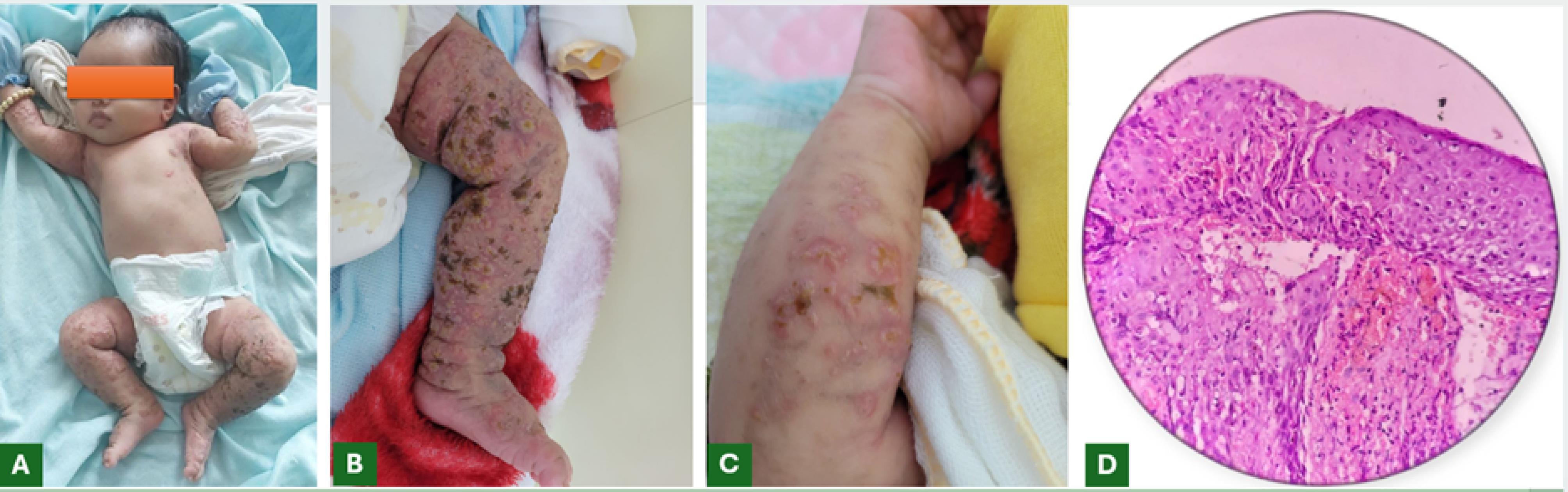
Figure 1.
(A, B, C) The clinical presentation revealed vesicles and tense bullae on an erythematous base, distributed along an apparent Blaschko-linear pattern predominantly involving the patient’s extremities; (D) The histopathology slide demonstrated prominent eosinophilic spongiosis with intraepidermal vesicles containing eosinophils.
.
(A, B, C) The clinical presentation revealed vesicles and tense bullae on an erythematous base, distributed along an apparent Blaschko-linear pattern predominantly involving the patient’s extremities; (D) The histopathology slide demonstrated prominent eosinophilic spongiosis with intraepidermal vesicles containing eosinophils.
The vesicular stage is the first stage of incontinentia pigmenti (IP), a disease with four distinct stages, each corresponding to the individual’s growth. The estimated incidence of IP is approximately 0.7 cases per 100 000 births.1 As an X-linked dominant genetic disorder, IP manifests predominantly in females because affected males typically cannot survive until birth. The characteristic clinical symptoms of vesicular stage IP include vesicles and tense bullae on an erythematous base, distributed along Blaschko lines.2 Diagnosing IP is generally straightforward but may pose challenges for less experienced physicians. When clinical assessment is inconclusive, supportive diagnostic tools include complete blood count demonstrating eosinophil-predominant leukocytosis and histopathological evidence of eosinophilic spongiosis. Family history evaluation is also crucial, particularly investigating dermatological conditions in female relatives on the maternal side of the patient’s family and documentation of miscarriages or absence of male offspring in the maternal lineage. In most cases, the diagnosis of vesicular stage IP still relies primarily on cutaneous manifestations.3 This principle is reflected in the 2014 diagnostic criteria,4 which require at least one major criterion, characteristic stage 1 skin findings, and at least one minor criterion, frequently histopathological evidence of eosinophilic spongiosis, particularly when extracutaneous involvement is relatively subtle or goes unnoticed in otherwise healthy infants like our case.
In summary, the triad of characteristic skin lesions, peripheral eosinophilia, and histopathological eosinophilic spongiosis can be considered the cornerstone for diagnosing the vesicular stage of IP.
Competing Interests
No conflict of interest to declare.
Ethical Approval
We obtained informed consent from the patient’s relatives to publish this case report.
Funding
The authors did not receive support from any organization for the submitted work.
References
- Cammarata-Scalisi F, Fusco F, Ursini MV. Incontinentia pigmenti. Actas Dermosifiliogr (Engl Ed) 2019; 110(4):273-8. doi: 10.1016/j.adengl.2019.03.009 [Crossref] [ Google Scholar]
- Çetinarslan T, Fölster-Holst R, Van Gysel D, Buchner M, Happle R. Incontinentia pigmenti stage 1 is not simply vesiculo-bullous but vesiculo-pustular. Pediatr Dermatol 2024; 41(1):182-3. doi: 10.1111/pde.15465 [Crossref] [ Google Scholar]
- Pal T, Agrawal S, Grover C. Incontinentia pigmenti. Indian Pediatr 2024; 61(8):799. doi: 10.1007/s13312-024-3268-z [Crossref] [ Google Scholar]
- Andrade A, Gonçalves CF, Fernandes PV, Jacinto T, Teixeira F, Costa E. Incontinentia pigmenti - one case, two diagnoses. J Clin Images Med Case Rep 2024; 5(2):2831. doi: 10.52768/2766-7820/2831 [Crossref] [ Google Scholar]